
Rod-Cone Dystrophy
Rod-cone dystrophy is a genetic condition that causes progressive vision loss, often starting in childhood.
Lattice degeneration is thinning of the retina’s outer edge marked by crisscross white lines; usually symptom-free but can create holes or tears that raise detachment risk.
Hear from Dr. Rosenfarb

Whether you prefer hands-on care, convenient telehealth visits, or self-guided learning, we have multiple ways to help you manage Lattice Degeneration.

Start here. A member of our care team will review your condition and situation, answer your questions, and walk you through the treatment options that are the best fit for you.
Book your free assessment call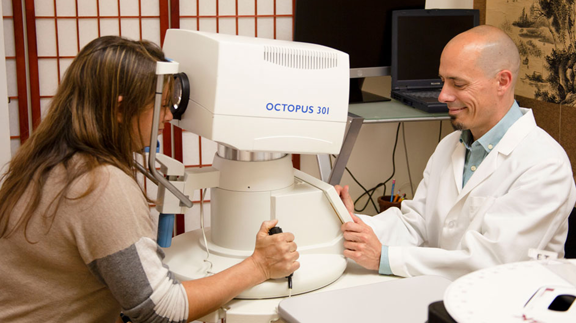
Combining acupuncture, laser therapy & diagnostics at Dr. Rosenfarb's office in New Jersey. 90% of patients see measurable vision improvements.
Learn more
One-on-one virtual sessions with Dr. Rosenfarb. Get personalized assessment and custom treatment plan from home.
Learn more
Scientifically-formulated supplements chosen by Dr. Rosenfarb to nourish your eyes and support healthy vision recovery.
Get supplementsDr. Rosenfarb's top-recommended supplements to nourish and protect your eyes.

Supplements
$65.00

Eye Drops & Lubricants
$40.00$35.00
Save 13%

Supplements
$70.00

Vitamins & Supplements
$30.00
Ready to take the next step?
Choose whatever feels right for you — no pressure, no commitment.
Rated 5 stars by 10,000+ Happy Patients Worldwide
A real patient shares their journey with our treatment approach.
"The eye floaters I had? They’re gone!"
Luxury real estate agent Lisa was urged to undergo surgery for lattice degeneration and a macular pucker. But after just two weeks on Dr. Rosenfarb’s integrative treatment plan, her floaters disappeared, night glare improved, and reading contracts became effortless. No scalpel, no downtime, no missed closings.
Common questions we get asked about Lattice Degeneration.
No. While lattice degeneration increases the chance of developing a retinal tear, most people never progress to a detachment, especially when they receive regular dilated eye exams and know the danger signs.
For uncomplicated lattice degeneration, an annual dilated retinal exam is typical. Your doctor may recommend a visit every 6 months if you are highly myopic, have new floaters or flashes, or have a strong family history of retinal detachment.
Barrier laser photocoagulation is usually done in the office with topical anesthetic drops. Most patients describe a brief mild stinging or warmth rather than true pain, and vision usually returns to normal within a few hours.
Protective eyewear for contact sports, good blood-sugar control, and avoiding activities that cause sudden head or eye jolts (like bungee jumping) can all help. A nutrient-rich diet with antioxidants and omega-3 fatty acids may also support overall retinal health.
The thinned retinal patches themselves do not re-thicken, but they often remain stable for life. The goal is to monitor for new holes or tears so they can be treated before a detachment develops.
There is no clear single-gene pattern, but lattice degeneration is more common in families with high myopia or connective-tissue disorders. If close relatives have had a retinal detachment, be extra vigilant with regular eye exams.
Discover other eye conditions that share similar causes, symptoms, or treatment approaches with the one you're exploring.
Rod-cone dystrophy is a genetic condition that causes progressive vision loss, often starting in childhood.
Stargardt syndrome is an inherited juvenile macular degeneration caused by gene mutations, leading to gradual central vision loss.
Usher syndrome combines inherited hearing loss with progressive night and peripheral vision decline from retinitis pigmentosa, often accompanied by balance problems.