Glaucoma / Optic Nerve
Glaucoma is a group of eye diseases in which elevated eye pressure damages the optic nerve, silently stealing peripheral vision and potentially leading to blindness.
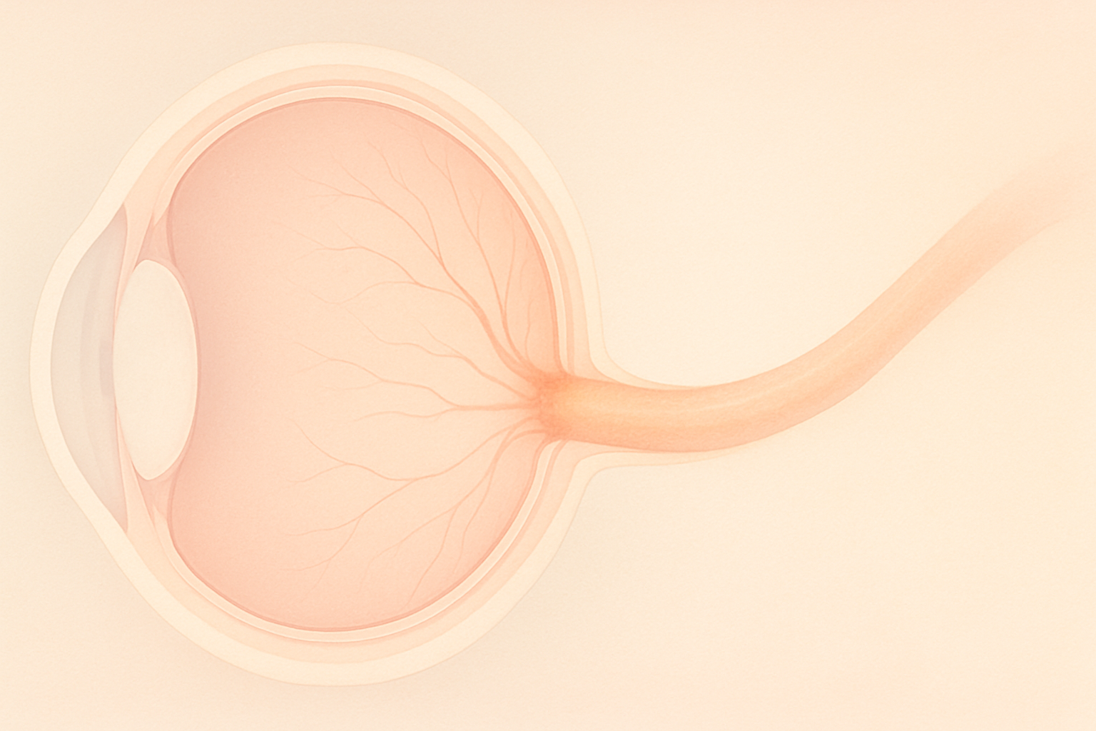
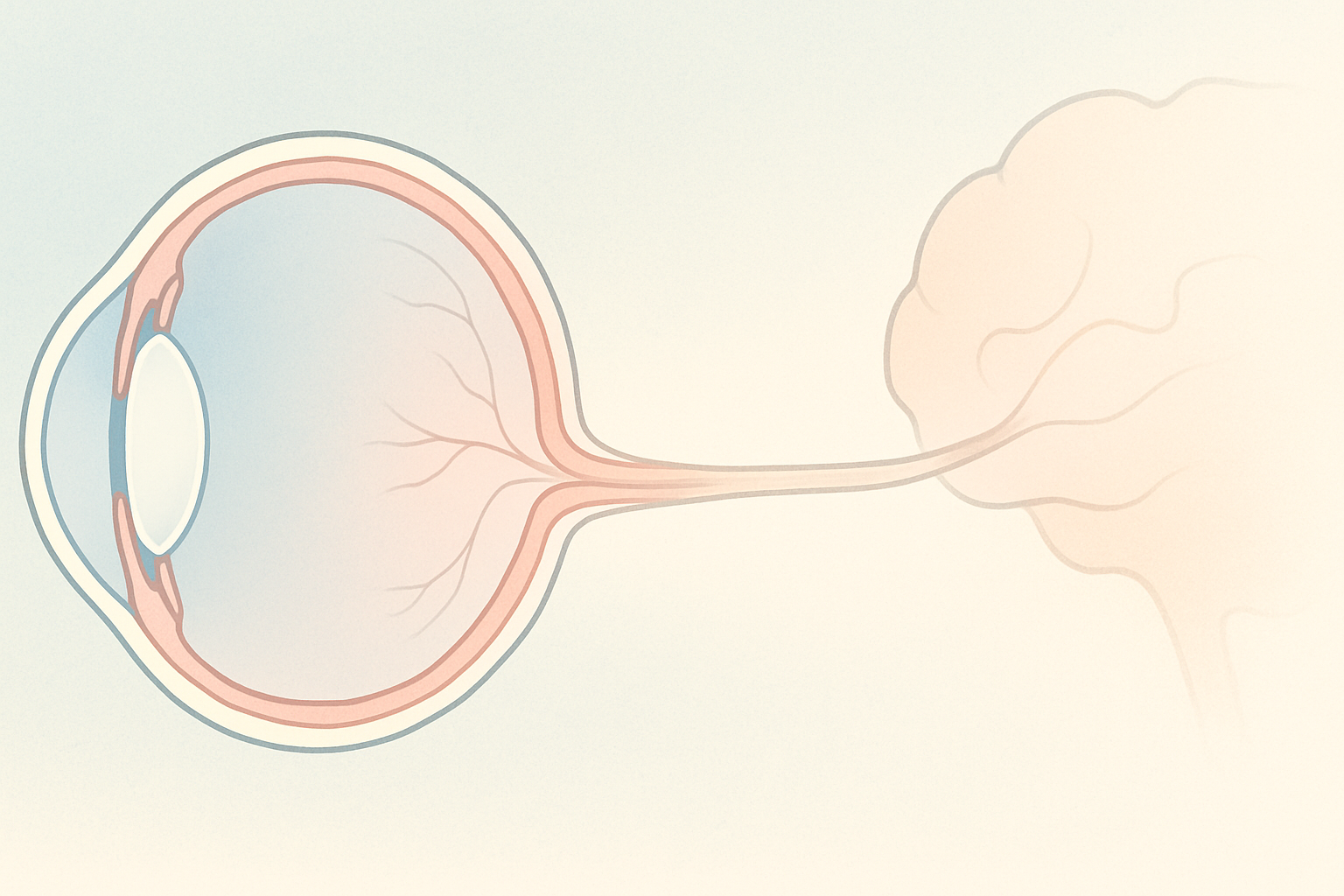
Optic nerve atrophy limits the optic nerve’s ability to relay visual signals, leading to progressive vision loss.
Hear from Dr. Rosenfarb
Whether you prefer hands-on care, convenient telehealth visits, or self-guided learning, we have multiple ways to help you manage Optic Nerve Atrophy.

Start here. A member of our care team will review your condition and situation, answer your questions, and walk you through the treatment options that are the best fit for you.
Book your free assessment call
Combining acupuncture, laser therapy & diagnostics at Dr. Rosenfarb's office in New Jersey. 90% of patients see measurable vision improvements.
Learn more
One-on-one virtual sessions with Dr. Rosenfarb. Get personalized assessment and custom treatment plan from home.
Learn more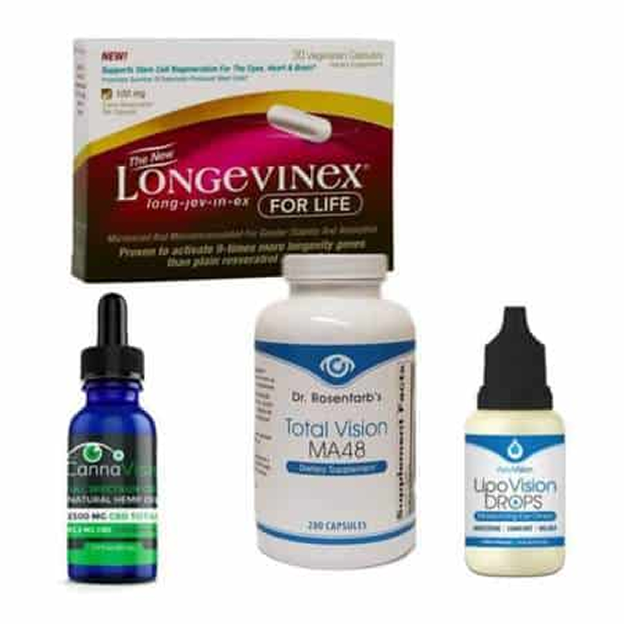
Scientifically-formulated supplements chosen by Dr. Rosenfarb to nourish your eyes and support healthy vision recovery.
Get supplementsDr. Rosenfarb's top-recommended supplements to nourish and protect your eyes.

Supplements
$65.00

Supplements
$70.00

Vitamins & Supplements
$30.00

Supplements
$30.00$25.00
Save 17%
Ready to take the next step?
Choose whatever feels right for you — no pressure, no commitment.
Rated 5 stars by 10,000+ Happy Patients Worldwide
A real patient shares their journey with our treatment approach.
"My peripheral vision is better and coming back. This is working for me."
Jim sought acupuncture at the Eye Health Institute after ischemic optic neuropathy left his right eye centrally blind. Since starting treatment, both his peripheral and central vision have improved, and he feels the therapy is genuinely helping.
Common questions we get asked about Optic Nerve Atrophy.
Yes. Some hereditary and metabolic forms appear in early childhood, while others surface during puberty. Regular pediatric eye exams are the best way to catch changes before they interfere with school or sports.
Pace varies by cause. A vascular event such as ischemic optic neuropathy may remove vision in hours, whereas genetic or compressive causes can unfold over months to years. Stabilizing the underlying trigger often slows or stops further loss.
Most people feel no eye pain at all. The exception is optic neuritis, an inflammatory cousin, which can cause soreness with eye movement. Pain should always prompt urgent evaluation to rule out treatable inflammation.
It depends on your visual-field results and local licensing rules. Many jurisdictions require at least 120° of horizontal field and a certain level of acuity. Low-vision specialists can test you and document whether you meet those standards.
They reduce glare and eye fatigue but have no proven effect on optic-nerve health. Protecting nerve tissue hinges more on blood flow, mitochondrial support, and controlling systemic inflammation.
If a doctor suspects a hereditary optic neuropathy,such as Leber hereditary optic neuropathy or dominant optic atrophy, a DNA panel can confirm the diagnosis, inform family planning, and guide monitoring of relatives.
Diets rich in leafy greens, omega-3-fatty-acid fish, nuts, and colorful berries provide B-vitamins, lutein, and antioxidants. Clinicians often add targeted nutrients such as vitamin B12, folate, alpha-lipoic acid, CoQ10, and omega-3 capsules when bloodwork shows a gap.
Coverage is inconsistent. Some plans reimburse medically coded acupuncture visits, but most consider microcurrent devices, red-light glasses, and nutraceuticals elective. Patients commonly use HSA or FSA funds to offset costs.
Discover other eye conditions that share similar causes, symptoms, or treatment approaches with the one you're exploring.
Glaucoma is a group of eye diseases in which elevated eye pressure damages the optic nerve, silently stealing peripheral vision and potentially leading to blindness.
Optic neuritis is an immune-driven inflammation of the optic nerve that can cause sudden vision loss, eye pain, and color desaturation, yet many people recover well with timely, integrative care.