Best's Disease
Best’s disease, also known as Best’s vitelliform macular dystrophy, is a hereditary (usually) form of progressive macular dystrophy.
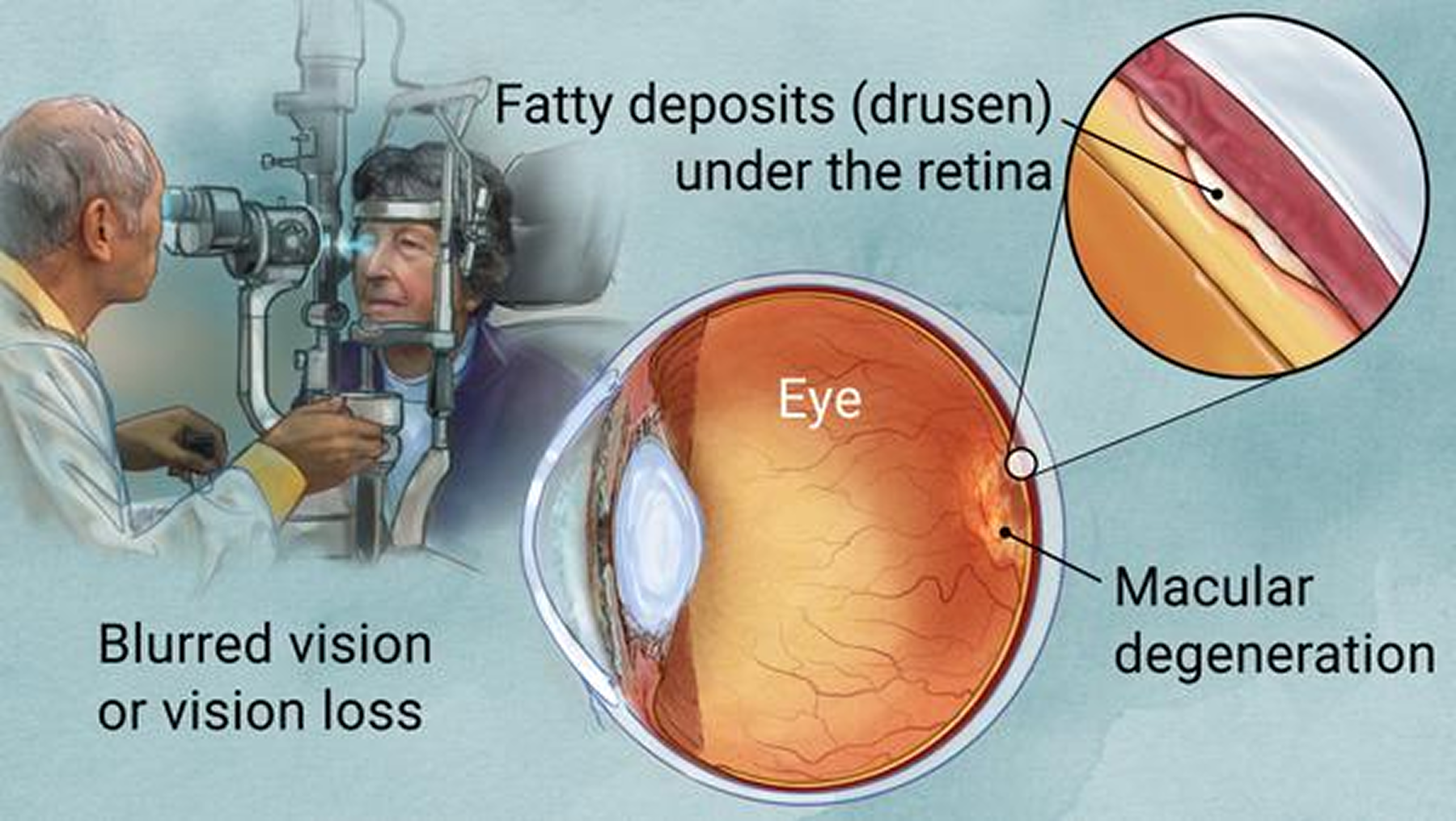
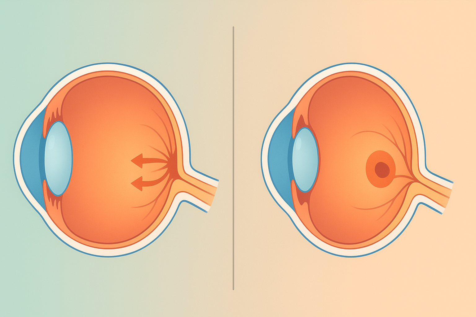
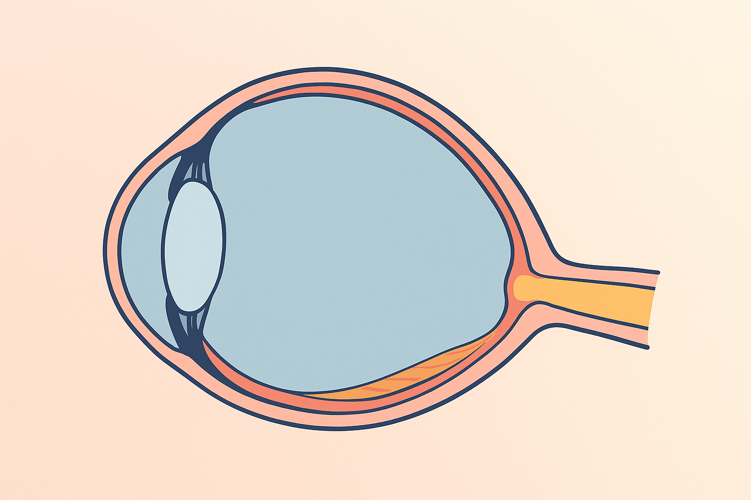
Wet macular degeneration arises when abnormal retinal blood vessels leak beneath the macula, causing rapid distortion and loss of central vision.
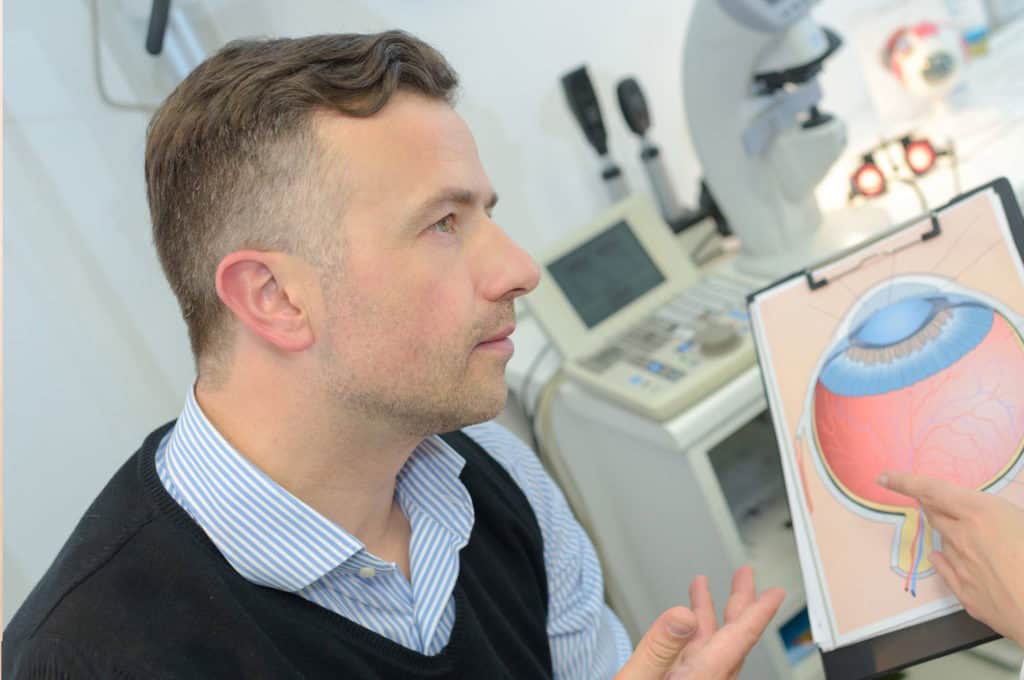
Hear from Dr. Rosenfarb
Whether you prefer hands-on care, convenient telehealth visits, or self-guided learning, we have multiple ways to help you manage Wet Macular Degeneration.

Start here. A member of our care team will review your condition and situation, answer your questions, and walk you through the treatment options that are the best fit for you.
Book your free assessment call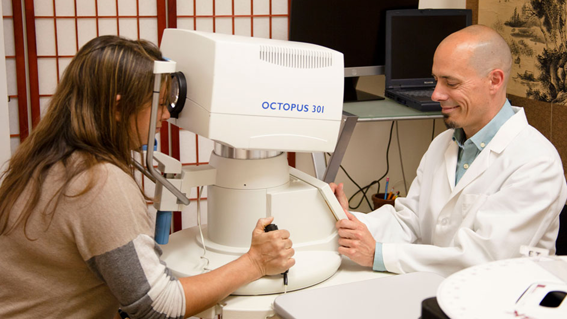
Combining acupuncture, laser therapy & diagnostics at Dr. Rosenfarb's office in New Jersey. 90% of patients see measurable vision improvements.
Learn more
One-on-one virtual sessions with Dr. Rosenfarb. Get personalized assessment and custom treatment plan from home.
Learn more
Scientifically-formulated supplements chosen by Dr. Rosenfarb to nourish your eyes and support healthy vision recovery.
Get supplementsDr. Rosenfarb's top-recommended supplements to nourish and protect your eyes.

Supplements
$65.00

Vitamins & Supplements
$30.00

Supplements
$30.00$25.00
Save 17%

Eye Drops & Lubricants
$50.00$39.99
Save 20%
Ready to take the next step?
Choose whatever feels right for you — no pressure, no commitment.
Rated 5 stars by 10,000+ Happy Patients Worldwide
A real patient shares their journey with our treatment approach.
"After electro-acupuncture, the little patterns are gone, like my retinal cells woke up!"
At 80, scientist Melissa battled wet AMD, geographic atrophy and Lyme‑linked Bartonella. In just three visits over a year, Dr. Rosenfarb’s electro‑acupuncture and hydrogen therapy erased disruptive visual patterns, re‑awoke dormant retinal cells, and steadied her vision—prompting her to urge others: “don’t wait.”
Common questions we get asked about Wet Macular Degeneration.
Dry AMD is a slow thinning of macular tissue, while wet AMD occurs when new, fragile blood vessels grow and leak beneath the retina, leading to quicker and more severe central‑vision loss.
The mainstay is anti‑VEGF medication injected into the eye every few weeks to stop vessel leakage; some patients also benefit from photodynamic therapy or, in select cases, focal laser treatment.
Before each injection your eye is numbed with anesthetic drops, so most people feel only mild pressure; the needle is ultra‑fine and the procedure is designed not to harm healthy eye structures.
Many patients start with monthly injections for the first 3 – 6 months; if the retina stays dry on scans, the interval can be gradually extended to every 8–12 weeks under your specialist’s guidance.
Quitting smoking, maintaining healthy blood pressure, and eating a diet rich in dark leafy greens, omega‑3 fatty acids, and antioxidant‑containing foods may help protect retinal cells and the other eye.
You may continue driving as long as you meet your state’s visual‑acuity requirements; regular eye exams are essential, and adaptive devices such as wrap‑around glare‑reducing lenses can improve safety.
Discover other eye conditions that share similar causes, symptoms, or treatment approaches with the one you're exploring.
Best’s disease, also known as Best’s vitelliform macular dystrophy, is a hereditary (usually) form of progressive macular dystrophy.
Central serous retinopathy is a condition that causes fluid to leak from the choroid layer into the macula, causing blurring or distortion of central vision.
Macular degeneration erodes the macula and central vision, but it’s often a sign of broader circulation, inflammation, and energy imbalances.
Macular dystrophy is a rare genetic disorder that slowly damages the macula, the eye's center for sharp vision, resulting in progressive central vision loss.
Macular edema, also called cystoid macular edema, is swelling in the retina’s center that blurs detail; our goal is early detection & integrative care to help protect sight.
Macular pucker (epiretinal membrane) is a thin scar layer on the macula that contracts, wrinkling the retina and blurring or distorting central vision.
Myopic degeneration is severe nearsightedness that stretches and thins eye tissues, causing progressive vision loss and higher retinal detachment risk.
Pattern dystrophy is an inherited retinal disorder in which pigment collects in distinctive macular patterns, slowly causing central vision to blur.