
Eye Floaters
Eye floaters are tiny drifting shapes caused by age-related changes in the eye’s vitreous gel, and while usually harmless they can sometimes signal retinal danger.
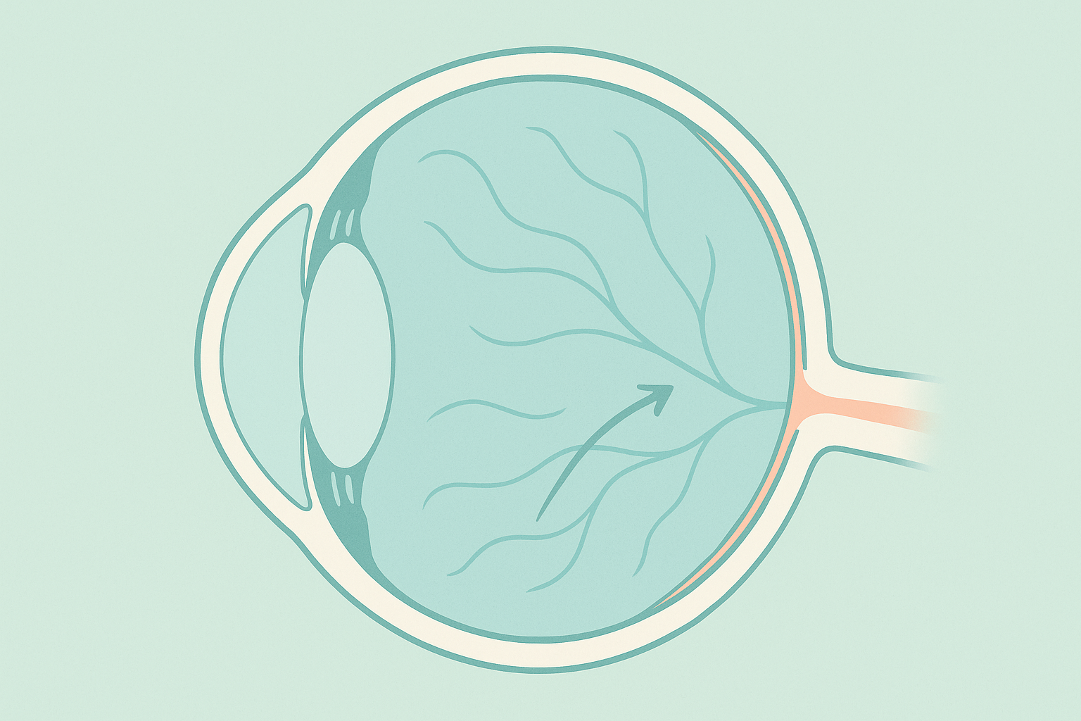
Vitreous detachment is a common eye condition that occurs when the vitreous gel, the clear jelly-like substance in the eye, separates from the retina.
Hear from Dr. Rosenfarb
Whether you prefer hands-on care, convenient telehealth visits, or self-guided learning, we have multiple ways to help you manage Vitreous Detachment.

Start here. A member of our care team will review your condition and situation, answer your questions, and walk you through the treatment options that are the best fit for you.
Book your free assessment call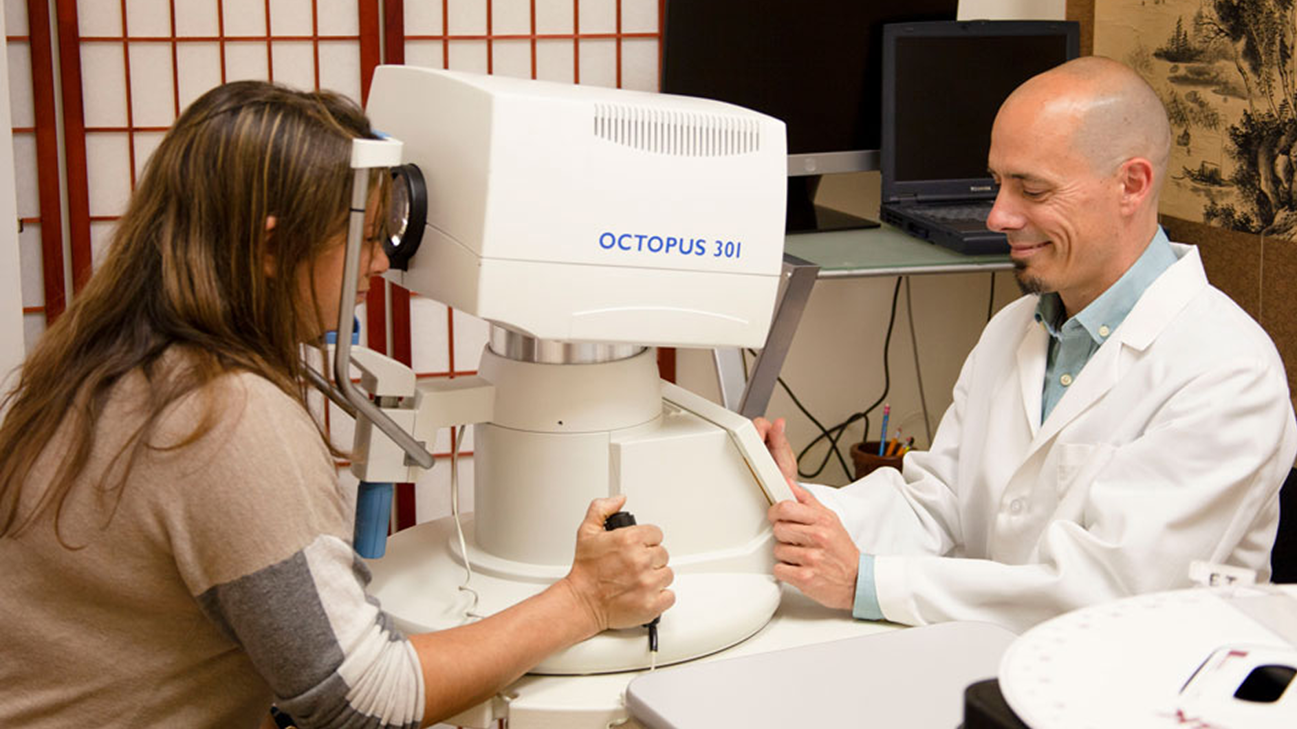
Combining acupuncture, laser therapy & diagnostics at Dr. Rosenfarb's office in New Jersey. 90% of patients see measurable vision improvements.
Learn more
One-on-one virtual sessions with Dr. Rosenfarb. Get personalized assessment and custom treatment plan from home.
Learn more
Scientifically-formulated supplements chosen by Dr. Rosenfarb to nourish your eyes and support healthy vision recovery.
Get supplementsDr. Rosenfarb's top-recommended supplements to nourish and protect your eyes.

Supplements
$65.00

Supplements
$70.00

Vitamins & Supplements
$30.00

Supplements
$30.00$25.00
Save 17%
Ready to take the next step?
Choose whatever feels right for you — no pressure, no commitment.
Common questions we get asked about Vitreous Detachment.
No. Vitreous detachment is the separation of the eye’s gel from the retina and is common with age, whereas retinal detachment is a serious condition where the retina itself peels away from the back of the eye. Vitreous detachment can raise the risk of a retinal tear, but the two are not identical.
For the first few weeks avoid high-impact sports, heavy weight-lifting, and sudden head movements that could increase tugging on the retina. Normal walking, desk work, and light household tasks are generally fine unless your eye doctor advises otherwise.
Screens do not change the floaters themselves, but the bright uniform background can make them more noticeable. Using a matte screen protector, lowering brightness, and taking regular blink breaks can reduce annoyance.
No supplement has been proven to dissolve existing floaters, but antioxidants such as vitamin C and collagen-supportive nutrients may promote overall vitreous and retinal health. Discuss any regimen with your eye-care professional.
Flying is usually safe once your ophthalmologist confirms there is no retinal tear. Night driving is allowed, but be prepared for glare from headlights if you have large floaters. Seek a prompt re-check if vision changes suddenly.
Most patients need a repeat dilated exam four to six weeks after the event, then annually unless new symptoms arise. Your doctor may tailor the schedule if you are highly nearsighted or have other retinal risk factors.
While aging is the main trigger, poorly controlled blood pressure and blood sugar can weaken retinal blood vessels and connective tissue, increasing the chance of complications. Managing systemic health is an important part of prevention.
Call or visit an emergency eye clinic immediately if you notice a sudden storm of new floaters, repeated bright flashes, a dark curtain in your vision, or any rapid loss of side or central vision. These symptoms can signal a retinal tear or detachment.
Discover other eye conditions that share similar causes, symptoms, or treatment approaches with the one you're exploring.
Eye floaters are tiny drifting shapes caused by age-related changes in the eye’s vitreous gel, and while usually harmless they can sometimes signal retinal danger.