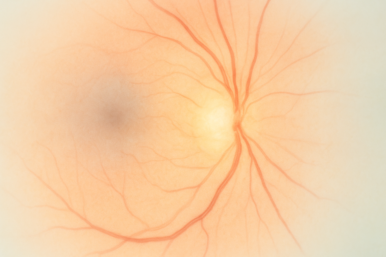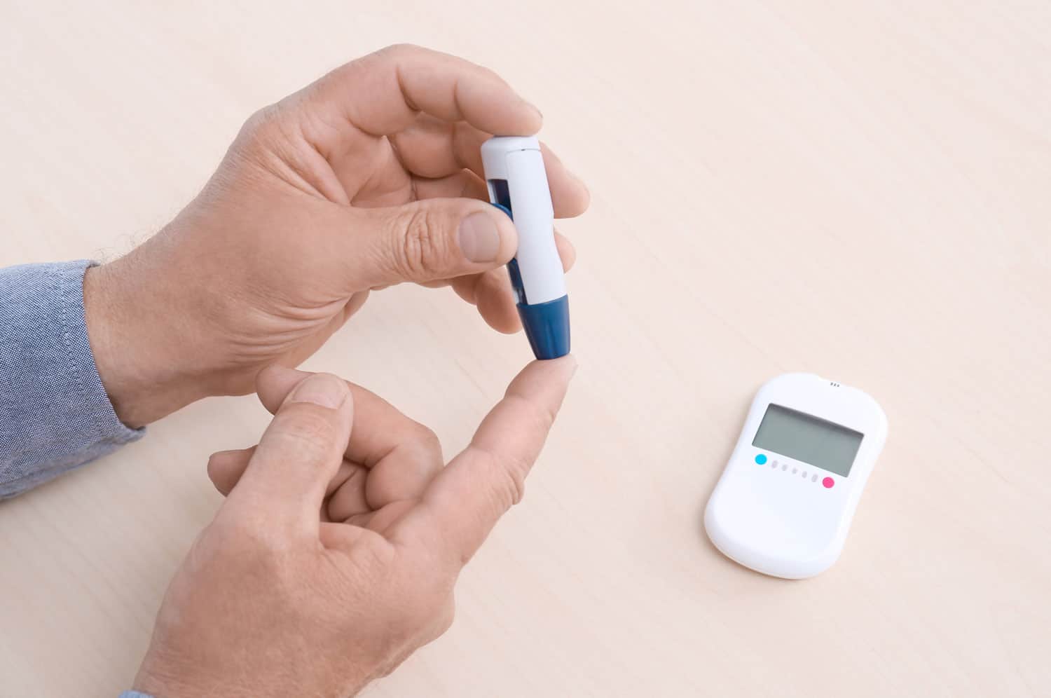
Diabetic Retinopathy
Diabetic retinopathy develops when high blood sugar damages retinal blood vessels, causing blurry vision, floaters, and potentially permanent vision loss.
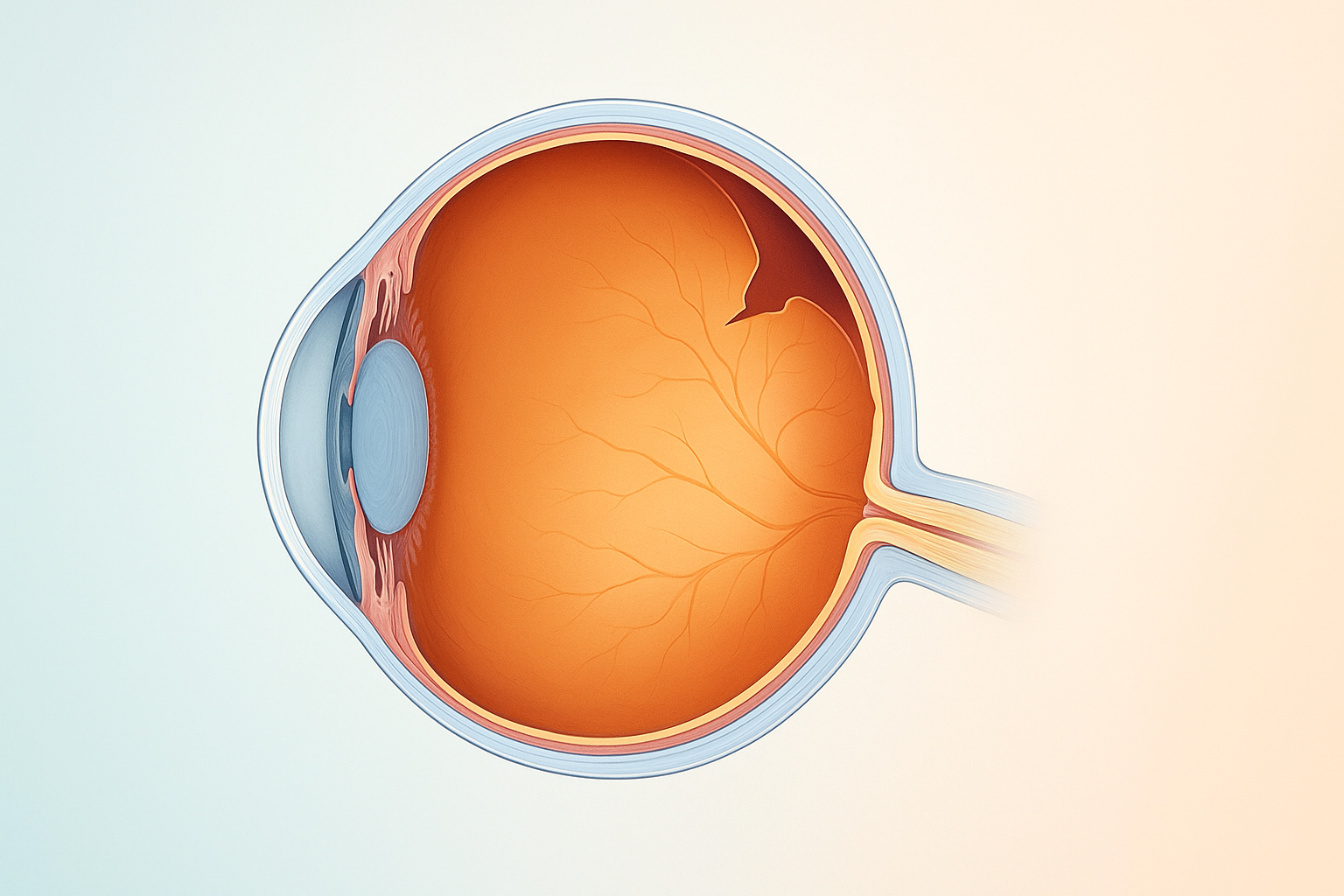
Retinal detachment is a medical emergency in which the light-sensitive retina peels away from the eye's back wall, triggering sudden flashes, floaters, and rapid vision loss.
Hear from Dr. Rosenfarb
Whether you prefer hands-on care, convenient telehealth visits, or self-guided learning, we have multiple ways to help you manage Retinal Detachment.

Start here. A member of our care team will review your condition and situation, answer your questions, and walk you through the treatment options that are the best fit for you.
Book your free assessment call
Combining acupuncture, laser therapy & diagnostics at Dr. Rosenfarb's office in New Jersey. 90% of patients see measurable vision improvements.
Learn more
One-on-one virtual sessions with Dr. Rosenfarb. Get personalized assessment and custom treatment plan from home.
Learn more
Scientifically-formulated supplements chosen by Dr. Rosenfarb to nourish your eyes and support healthy vision recovery.
Get supplementsDr. Rosenfarb's top-recommended supplements to nourish and protect your eyes.

Supplements
$65.00

Supplements
$70.00

Vitamins & Supplements
$30.00

Supplements
$30.00$25.00
Save 17%
Ready to take the next step?
Choose whatever feels right for you — no pressure, no commitment.
Rated 5 stars by 10,000+ Happy Patients Worldwide
A real patient shares their journey with our treatment approach.
"Every time I’ve come here, the colors seem way brighter for many months after the treatment."
Retinal tears in her teens left Mariana with floaters, flashes, and washed-out color. After a week of micro-acupuncture her right-eye field opened by 30%, the flashes calmed, and colors came alive again, results so encouraging she says she will happily return every year for the rest of her life.
Common questions we get asked about Retinal Detachment.
Yes. If you have had a detachment in one eye, the risk in the fellow eye is several times higher than average, so regular dilated exams are strongly advised.
Most detachments are painless; the danger comes from sudden visual changes such as new floaters, flashes, or a shadow that moves across your vision.
Visual stabilization can take weeks to months, but most people resume light activities within 2–4 weeks, depending on the surgical method and your doctor’s instructions.
No. Air travel (and any change in altitude) must be avoided until the injected gas bubble is fully absorbed, because pressure changes can dangerously expand the bubble.
Re‑detachment is possible, especially within the first few months; prompt follow‑up appointments let your surgeon catch and treat any new problems early.
Follow precise head‑positioning instructions, avoid strenuous exercise, protect the eye from impact, and stick to all eye‑drop schedules to reduce inflammation and infection risk.
Discover other eye conditions that share similar causes, symptoms, or treatment approaches with the one you're exploring.
Diabetic retinopathy develops when high blood sugar damages retinal blood vessels, causing blurry vision, floaters, and potentially permanent vision loss.
NAION and retinal occlusion (eye stroke) both cut off blood flow to the eye — one to the optic nerve, the other to the retina. Both demand urgent integrative care to protect and restore vision.
A retinal tear is a small rupture in the eye’s retina that can trigger sudden flashes or floaters and may progress to detachment if untreated.
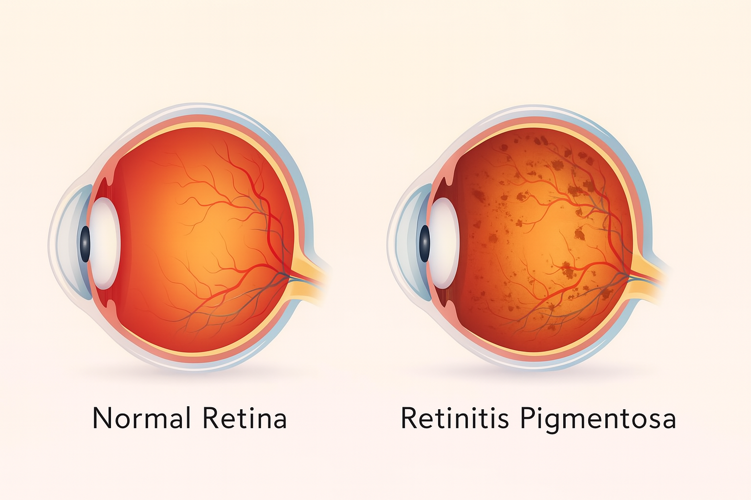
Retinitis pigmentosa is an inherited retinal disorder that gradually destroys photoreceptor cells, leading to night blindness and progressive tunnel vision.