Central Serous Retinopathy
Central serous retinopathy is a condition that causes fluid to leak from the choroid layer into the macula, causing blurring or distortion of central vision.
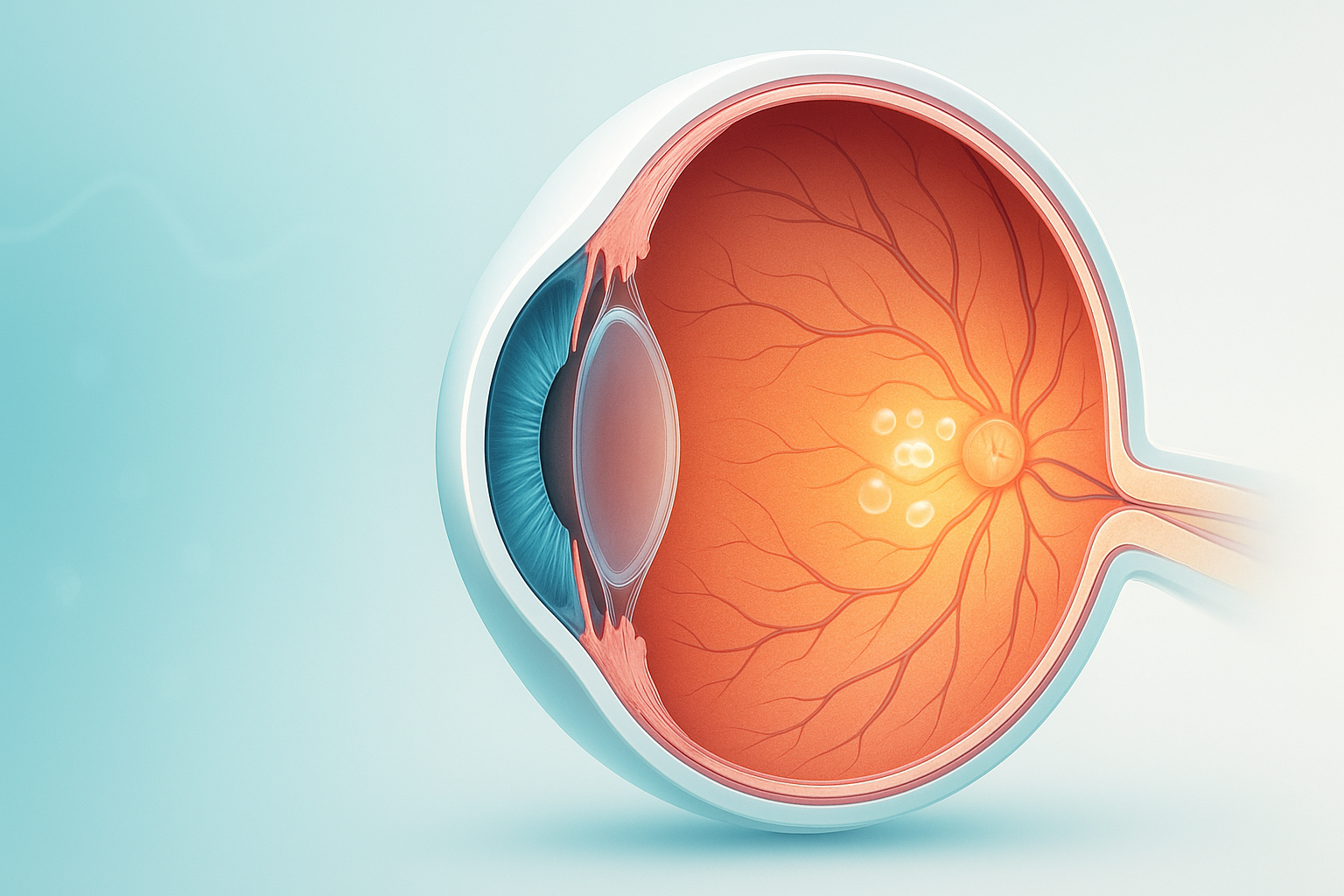
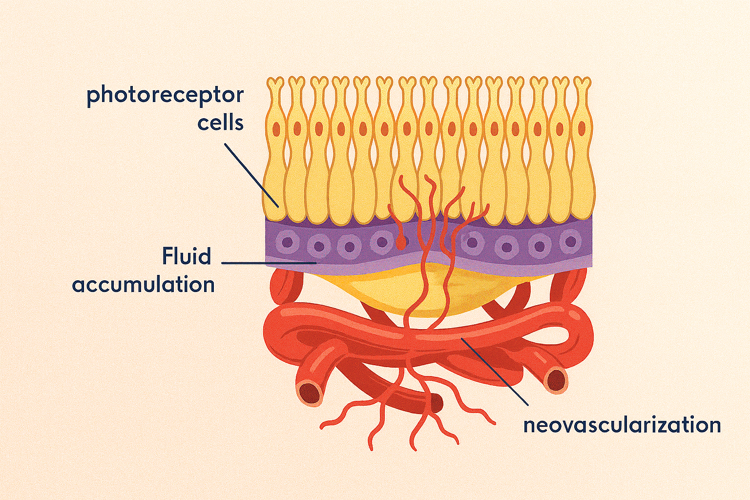
Best’s disease, also known as Best’s vitelliform macular dystrophy, is a hereditary (usually) form of progressive macular dystrophy.
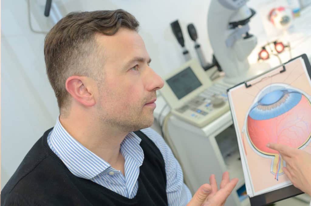
Hear from Dr. Rosenfarb
Whether you prefer hands-on care, convenient telehealth visits, or self-guided learning, we have multiple ways to help you manage Best's Disease.

Start here. A member of our care team will review your condition and situation, answer your questions, and walk you through the treatment options that are the best fit for you.
Book your free assessment call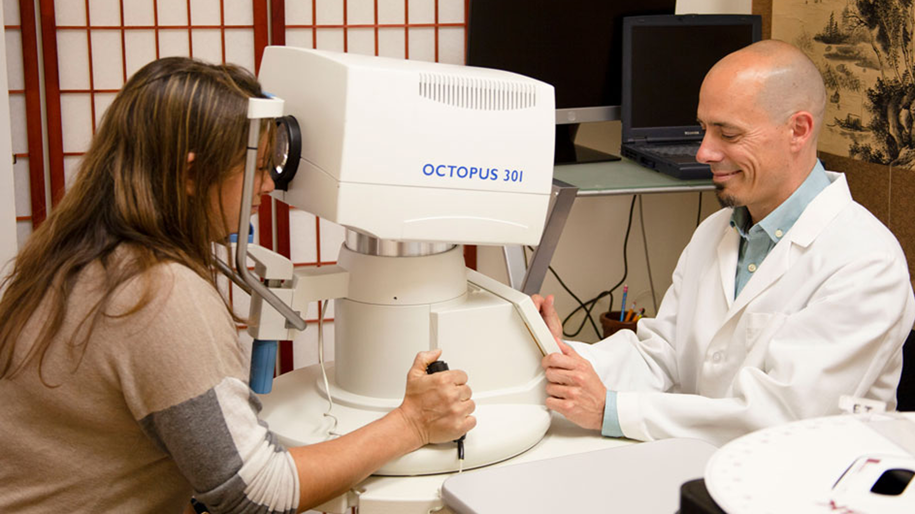
Combining acupuncture, laser therapy & diagnostics at Dr. Rosenfarb's office in New Jersey. 90% of patients see measurable vision improvements.
Learn more
One-on-one virtual sessions with Dr. Rosenfarb. Get personalized assessment and custom treatment plan from home.
Learn more
Scientifically-formulated supplements chosen by Dr. Rosenfarb to nourish your eyes and support healthy vision recovery.
Get supplementsDr. Rosenfarb's top-recommended supplements to nourish and protect your eyes.

Supplements
$65.00

Supplements
$70.00

Vitamins & Supplements
$30.00

Supplements
$30.00$25.00
Save 17%
Ready to take the next step?
Choose whatever feels right for you — no pressure, no commitment.
Rated 5 stars by 10,000+ Happy Patients Worldwide
A real patient shares their journey with our treatment approach.
"I have maintained my eyesight for over 30 years now."
Diagnosed in 1993 with Best disease and told she’d be blind in years, Janet turned to Dr. Rosenfarb’s acupuncture and hyperbaric oxygen. Three decades later she still drives, sees faces, and her macular lesions have even regressed, stunning her retinal surgeon.
Common questions we get asked about Best's Disease.
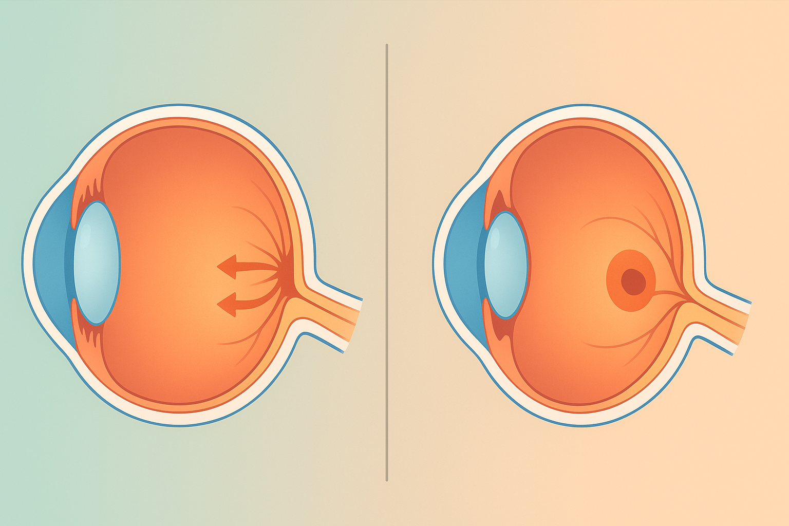
Both conditions damage the macula, but Best’s disease is inherited and usually begins in childhood, whereas age‑related macular degeneration develops later in life and is linked to aging rather than a single gene mutation.
Best’s disease typically follows an autosomal‑dominant pattern, so each child has a 50 percent chance of inheriting the gene and eventually developing some form of the condition.
Not always. Some carriers have normal vision and only subtle changes on specialized retinal testing, this is called reduced penetrance.
Unexplained drop in reading vision, a small central blur that doesn’t clear with blinking, or a yellowish spot seen during a routine eye screening are all reasons to seek a specialist’s opinion.
Most doctors recommend a detailed retinal exam and OCT imaging once or twice a year, increasing the frequency if new symptoms appear.
High‑resolution OCT can detect subtle changes in the photoreceptor layer and may help forecast which eyes are at higher risk of later atrophy or scarring.
Good overall eye health, balanced nutrition, avoiding smoking, wearing UV‑blocking sunglasses, may support retinal cells, but it does not alter the genetic cause of Best’s disease.
Yes. A genetic counselor can clarify inheritance patterns, discuss reproductive options, and connect families with emerging clinical trials or registries.
Discover other eye conditions that share similar causes, symptoms, or treatment approaches with the one you're exploring.
Central serous retinopathy is a condition that causes fluid to leak from the choroid layer into the macula, causing blurring or distortion of central vision.
Macular degeneration erodes the macula and central vision, but it’s often a sign of broader circulation, inflammation, and energy imbalances.
Macular dystrophy is a rare genetic disorder that slowly damages the macula, the eye's center for sharp vision, resulting in progressive central vision loss.
Macular edema, also called cystoid macular edema, is swelling in the retina’s center that blurs detail; our goal is early detection & integrative care to help protect sight.
Macular pucker (epiretinal membrane) is a thin scar layer on the macula that contracts, wrinkling the retina and blurring or distorting central vision.
Myopic degeneration is severe nearsightedness that stretches and thins eye tissues, causing progressive vision loss and higher retinal detachment risk.
Pattern dystrophy is an inherited retinal disorder in which pigment collects in distinctive macular patterns, slowly causing central vision to blur.
Wet macular degeneration arises when abnormal retinal blood vessels leak beneath the macula, causing rapid distortion and loss of central vision.