Best's Disease
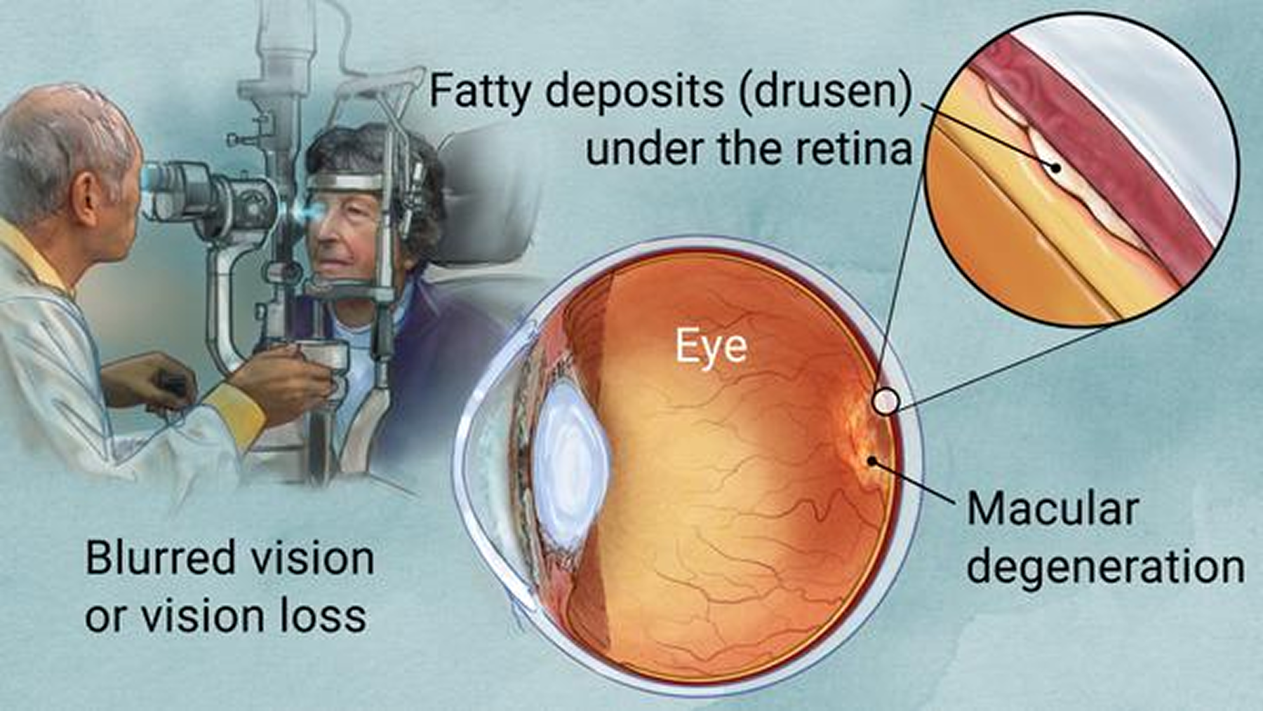
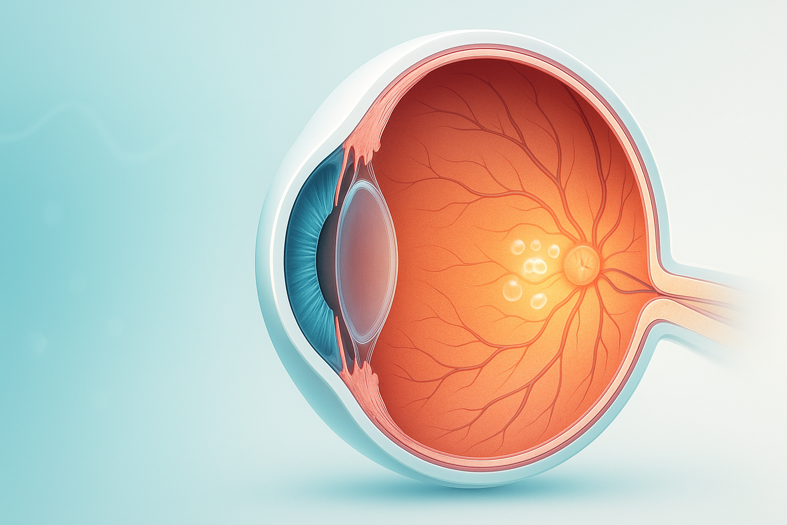
Best’s disease, also known as Best’s vitelliform macular dystrophy, is a hereditary (usually) form of progressive macular dystrophy.
Macular dystrophy is a rare genetic disorder that slowly damages the macula, the eye's center for sharp vision, resulting in progressive central vision loss.
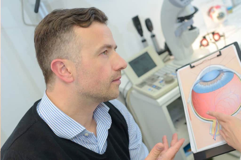
Hear from Dr. Rosenfarb
Whether you prefer hands-on care, convenient telehealth visits, or self-guided learning, we have multiple ways to help you manage Macular Dystrophy.

Start here. A member of our care team will review your condition and situation, answer your questions, and walk you through the treatment options that are the best fit for you.
Book your free assessment call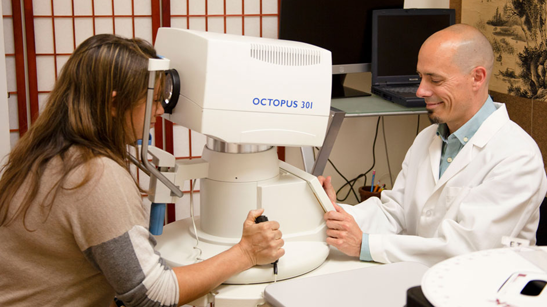
Combining acupuncture, laser therapy & diagnostics at Dr. Rosenfarb's office in New Jersey. 90% of patients see measurable vision improvements.
Learn more
One-on-one virtual sessions with Dr. Rosenfarb. Get personalized assessment and custom treatment plan from home.
Learn more
Scientifically-formulated supplements chosen by Dr. Rosenfarb to nourish your eyes and support healthy vision recovery.
Get supplementsDr. Rosenfarb's top-recommended supplements to nourish and protect your eyes.

Supplements
$65.00

Supplements
$70.00

Vitamins & Supplements
$30.00

Supplements
$30.00$25.00
Save 17%
Ready to take the next step?
Choose whatever feels right for you — no pressure, no commitment.
Common questions we get asked about Macular Dystrophy.
No. While both affect the macula, macular dystrophy is an inherited disorder that can appear earlier in life, whereas AMD is linked to aging and environmental factors.
There is currently no cure that restores lost macular cells, but early diagnosis, low‑vision aids, and emerging gene‑based treatments can help preserve remaining sight and quality of life.
Not necessarily, inheritance patterns vary. Genetic counseling can estimate each child’s risk and explain testing options.
A balanced diet rich in antioxidants supports overall retinal health, but no specific supplement has been proven to halt genetic macular dystrophy. Your doctor may still recommend nutrients that benefit eye tissue.
Most specialists advise a comprehensive retinal exam every 6–12 months, or sooner if you notice sudden vision changes.
That depends on how much central vision you retain and local licensing rules. Regular vision tests and adaptive driving aids can help many patients stay on the road safely.
High‑contrast smartphone settings, screen‑reader software, electronic magnifiers, and large‑print materials are popular tools that boost independence when central vision declines.
Discover other eye conditions that share similar causes, symptoms, or treatment approaches with the one you're exploring.
Best’s disease, also known as Best’s vitelliform macular dystrophy, is a hereditary (usually) form of progressive macular dystrophy.
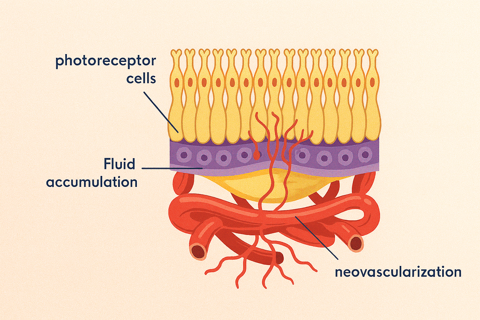
Central serous retinopathy is a condition that causes fluid to leak from the choroid layer into the macula, causing blurring or distortion of central vision.
Macular degeneration erodes the macula and central vision, but it’s often a sign of broader circulation, inflammation, and energy imbalances.
Macular edema, also called cystoid macular edema, is swelling in the retina’s center that blurs detail; our goal is early detection & integrative care to help protect sight.
Macular pucker (epiretinal membrane) is a thin scar layer on the macula that contracts, wrinkling the retina and blurring or distorting central vision.
Myopic degeneration is severe nearsightedness that stretches and thins eye tissues, causing progressive vision loss and higher retinal detachment risk.
Pattern dystrophy is an inherited retinal disorder in which pigment collects in distinctive macular patterns, slowly causing central vision to blur.
Wet macular degeneration arises when abnormal retinal blood vessels leak beneath the macula, causing rapid distortion and loss of central vision.